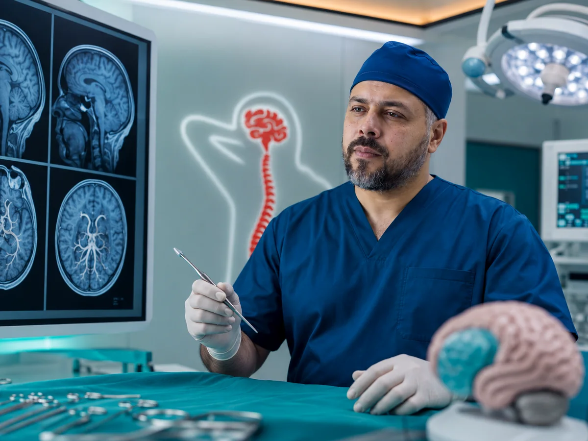
Hydrocephalus treatment starts with one question: why has the fluid built up? The answer decides whether a shunt that diverts the excess fluid, an endoscopic opening that creates a new pathway inside the brain, or treatment of the underlying cause is the right option. Prof. Mohamed El-Gaidi, Professor of Neurosurgery, treats hydrocephalus in adults and children at his clinic in Dokki, Cairo, and follows up patients with shunts.
What is hydrocephalus?
The brain continuously produces cerebrospinal fluid inside cavities called ventricles. The fluid circulates around the brain and spinal cord and is absorbed into the blood. When its pathway is blocked or absorption fails, fluid accumulates, the ventricles enlarge and pressure on the brain rises. Causes include congenital narrowing of the aqueduct, tumors or cysts, brain hemorrhage, previous meningitis and birth defects such as spina bifida; in some older adults no clear cause is found.
Types of hydrocephalus
Obstructive
Fluid is trapped by a blockage at a specific point, such as aqueductal stenosis, a posterior fossa tumor or a ventricular cyst. The best candidates for ETV.
Communicating
Pathways are open but absorption is poor, often after subarachnoid hemorrhage or meningitis. Usually needs a shunt.
Normal pressure (NPH)
Usually after 60. Ventricles enlarge without obvious headache, and symptoms develop slowly in walking, memory and bladder control.
A slow, shuffling gait as if the feet are stuck to the floor, forgetfulness and slowed thinking, and urinary urgency or incontinence. When these appear in an older person with enlarged ventricles on MRI, a surgical assessment is worthwhile: NPH is one of the treatable causes of "dementia".
Symptoms in children vs adults
| Infants and children | Adults |
|---|---|
| Head circumference growing faster than normal | Headache, worse in the morning or lying down |
| Bulging, tense fontanelle | Nausea and vomiting |
| Eyes fixed downward ("sunset eyes") | Blurred or double vision |
| Irritability, poor feeding, vomiting | Drowsiness and slowed responses |
| Delayed sitting, walking or speech | Walking and balance problems, memory decline |
For congenital conditions in children, see pediatric neurosurgery in Cairo.

How hydrocephalus is diagnosed
CT or MRI measures the ventricles and looks for the site and cause of the blockage; MRI shows the aqueduct, tumors and cysts in more detail. In infants, ultrasound through the fontanelle can be used for follow-up. An eye examination may look for optic disc swelling. In suspected NPH a lumbar tap test removes some fluid and walking is compared before and after; clear improvement favors benefit from a shunt.
NPH overlaps with Alzheimer's disease, Parkinson's disease and lumbar spinal stenosis, and may coexist with them. The best candidates are patients whose walking problem came first and dominates, whose symptoms are of shorter duration, and who improve after the tap test. Long-standing severe memory loss responds less, and you will hear this honestly before surgery.
Shunt or endoscopic third ventriculostomy?
A shunt is a thin tube under the skin with a valve that regulates drainage of excess fluid to another body cavity. An endoscopic third ventriculostomy (ETV) passes an endoscope through a small burr hole and makes an opening in the floor of the third ventricle so fluid bypasses the blockage.
- Ventriculoperitoneal (VP) shunt: ventricle to abdomen; the most widely used.
- Ventriculoatrial (VA) shunt: ventricle to the heart via a neck vein, when the abdomen is unsuitable.
- Lumboperitoneal (LP) shunt: lower back to abdomen, for selected communicating cases only.
| Comparison | Shunt (VP / VA / LP) | ETV |
|---|---|---|
| Suitable for | Most types, including communicating and NPH | Mainly obstructive, e.g. aqueductal stenosis |
| Implant | Yes, lifelong | No |
| Possible problems | Blockage, infection, overdrainage; may need revision | The opening can close; most failures occur in the first months |
| Age | All ages | Lower success in very young infants |
| Follow-up | Regular, lifelong | Close in the first year, then periodic |
Intraventricular endoscopy is also used to biopsy some ventricular tumors or open cysts that block fluid flow. When a tumor causes the hydrocephalus, it is treated as part of brain tumor surgery.
The type of hydrocephalus, its cause and the patient's age are the three factors that choose the route.
What happens during the operation
Both procedures are done under general anesthesia. For a shunt, a small burr hole lets the catheter enter the ventricle, and the tube is tunneled under the skin behind the ear, down the neck and chest to the abdomen through small incisions. For ETV, a thin endoscope enters through a single burr hole and nothing is left behind. Most patients stay a few days; headache and vomiting often improve quickly, while walking and memory in NPH improve gradually over weeks to months.
Warning signs of shunt malfunction
Go to the emergency department for returning severe headache with vomiting, drowsiness or difficulty waking, blurred vision, seizures, a bulging fontanelle and irritability in an infant, or redness and swelling along the shunt with fever or abdominal pain.
Headache that worsens on standing and eases lying down may mean overdrainage and needs a review with your surgeon, not just painkillers. Day to day, most people with shunts go to school, work and travel normally; airport scanners and phones do not affect them. Tell any surgeon operating on your abdomen that you have a shunt.
Follow-up after hydrocephalus treatment
Baseline scan
A scan soon after treatment becomes the reference for any later comparison.
Regular visits
Checking symptoms, walking and memory in adults; head size and development in children.
Valve adjustment
Some valves are programmable externally and are adjusted to the response and after any MRI.
Shunt card
Keep the valve type, date of surgery and latest scan to show any doctor or emergency team.
Your hydrocephalus surgeon in Dokki

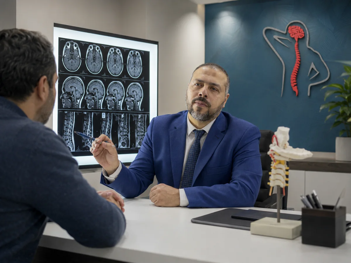
Hydrocephalus needs a surgeon who can choose between a shunt and endoscopy and then follow you for years. The clinic is at 114 El-Tahrir St., Dokki, Giza, next to Dokki Metro station, and receives patients from Cairo, Giza, other governorates and abroad. Bring all previous scans, operative notes and shunt details. If hydrocephalus followed a brain bleed, read about subarachnoid hemorrhage and brain aneurysms, or browse all neurosurgery services.