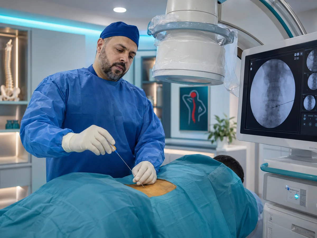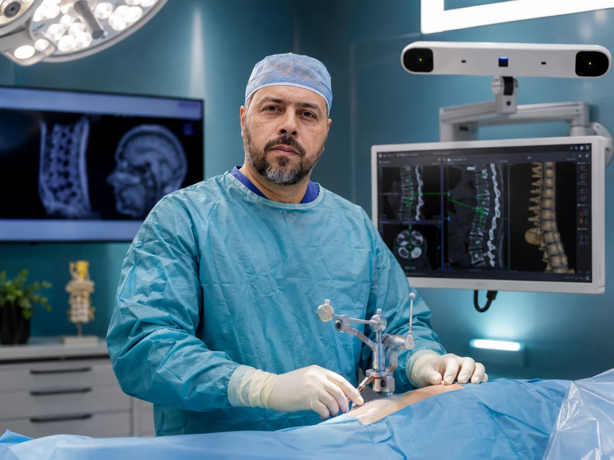
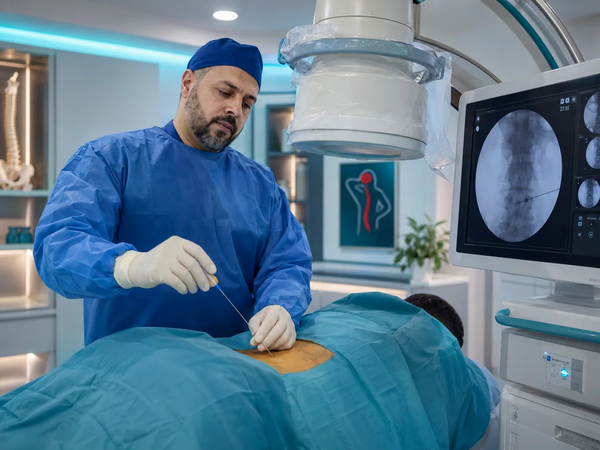
Radiofrequency ablation is a procedure in which an X-ray-guided needle is placed next to the small nerve that carries pain from a spinal joint, the knee or another joint, and a high-frequency current switches that pain signal off for a long time. It suits people with chronic pain that medication and physiotherapy have not controlled, and it needs no incision or stitches.
At Prof. Mohamed El-Gaidi's clinic in Dokki, Cairo, radiofrequency is part of a broader plan for back and neck pain treatment without surgery, and it is only performed once the pain source is confirmed. At your consultation you will learn whether conventional or pulsed radiofrequency fits your case.
How does radiofrequency stop pain?
Every joint has small sensory nerves that report what it feels to the brain. When the joint becomes arthritic, these nerves send constant pain with each movement. They carry sensation only and do not power the main muscles of the leg or arm, so they can be switched off without affecting walking or strength.
An insulated needle with an active tip is placed beside the nerve, its position is checked on X-ray and with a mild electrical test, and the generator then produces controlled heat at the tip only. The nerve stops transmitting pain while the joint and muscles stay as they are.
Conventional vs pulsed radiofrequency
There are two main types, chosen according to the nerve and the source of pain:
| Feature | Conventional radiofrequency | Pulsed radiofrequency |
|---|---|---|
| How it works | Continuous heat around 80°C creates a lesion in the small sensory nerve | Short bursts keep the temperature at about 42°C or below, modulating the pain signal without destroying the nerve |
| Target nerves | Sensory joint nerves: facet joints, sacroiliac joint, knee, hip | Nerve roots and mixed nerves that must not be damaged, such as the sciatic nerve root or the shoulder nerve |
| Typical use | Facet arthritis in the back and neck, chronic knee pain | Chronic sciatica and radicular pain, some shoulder and head pain |
| Duration of relief | Usually longer, several months or more | More variable; may be repeated |
| After the session | Soreness or burning for days to a few weeks | Usually milder |
A third option for selected knee, shoulder and hip pain is cryotherapy, which cools the nerve instead of heating it.
The diagnostic block comes first
Before conventional radiofrequency, a diagnostic block with local anaesthetic is placed on the same nerves. The logic is simple: if your pain disappears or clearly eases during the hours the anaesthetic works, those nerves are carrying the pain, and switching them off with radiofrequency should give similar relief for much longer.
After the block you record your pain and mobility through the day. A clear result means a radiofrequency session is scheduled. No improvement means the source lies elsewhere, and you have avoided a procedure that would not help. This step does more than anything else to raise the chance of success.
your pain has lasted more than three months, medication and physiotherapy have not been enough, your exam and scans point to a specific source, and a diagnostic block gave you clear relief.
Uses: back, neck and joint radiofrequency
Lumbar facet joints
Chronic low back pain that worsens when leaning back, twisting or standing for long, caused by arthritis of the small joints at the back of the spine.
Neck radiofrequency
Chronic neck pain spreading to the shoulders or the back of the head from cervical facet arthritis, particularly after old neck injuries.
Sacroiliac joint
One-sided low back pain above the buttock, treated by heating the small nerves that run from the sacrum to this joint.
Knee, shoulder and hip
Radiofrequency of the knee nerves eases chronic arthritis pain. For the shoulder and hip the joint's sensory nerves are targeted, and pulsed mode is often used at the shoulder.
Pulsed radiofrequency for sciatica
Leg pain spreading from the lower back (sciatica) usually comes from a compressed or inflamed nerve root. A nerve root carries both motor and sensory fibres, so conventional heat is not used there. Pulsed radiofrequency delivers gentle pulses to the root's ganglion, quietening the pain signal without affecting leg strength.
It is typically discussed when leg pain persists despite epidurals and physiotherapy, with no progressive weakness needing surgery, or when steroid injections are unsuitable. Diagnosis and the full treatment pathway are covered under sciatica treatment in Cairo, and if a large disc is the cause, see herniated disc treatment.
Radiofrequency for trigeminal and occipital nerve pain
Trigeminal neuralgia causes brief electric-shock attacks in the cheek, jaw or forehead, often triggered by eating, talking or touching the face. When medication is no longer enough or causes exhausting side effects, a needle can be guided by X-ray to the trigeminal branches and treated with radiofrequency. Some numbness in part of the face may remain afterwards, and this is explained in detail beforehand.
One-sided headache at the back of the head spreading over the scalp may come from the occipital nerve. After a positive diagnostic block, pulsed radiofrequency can reduce the attacks for longer.

What happens during a radiofrequency session?
Preparation
Blood thinners and diabetes medication are reviewed beforehand. On the day you lie on the table, the skin is cleaned and numbed, with light sedation if needed.
Needle guidance
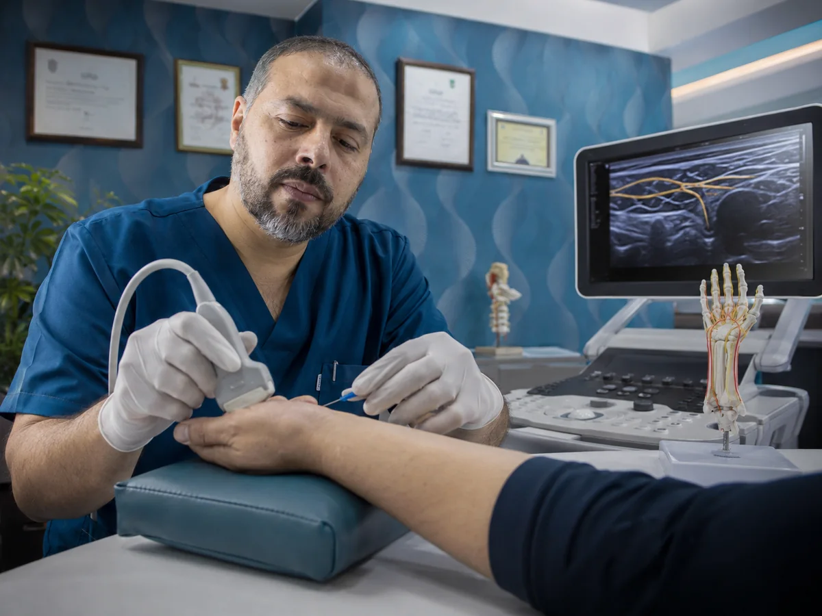
The needle is steered by X-ray, or by ultrasound for some joints, to the exact position of the nerve.
Electrical testing
A mild current confirms the needle sits beside the sensory nerve and away from motor nerves. You may feel a familiar tingling in your pain area.
Treatment and home
Local anaesthetic is injected around the nerve, then each level is treated. A session usually takes 30 to 60 minutes, and you go home afterwards.
How long does radiofrequency ablation last?
Full relief is not felt on day one. After conventional radiofrequency, needle-site pain may increase for a few days, then improvement builds gradually over roughly two to three weeks.
How long relief lasts differs between patients and joints. Many patients with facet arthritis stay better for several months, sometimes a year or longer. The small nerves regrow slowly, so if pain returns the procedure can usually be repeated after re-assessment. The effect of pulsed radiofrequency is more variable.
The realistic goal of radiofrequency is to move, sleep and work with less pain, and to use that window to strengthen the muscles around your spine or joint.
Side effects and who should not have it
Radiofrequency is generally safe when performed with proper guidance. The most common effects are:
- Soreness or bruising at the needle site for a few days.
- Burning or skin sensitivity over the treated area, usually settling within a few weeks.
- A small patch of numb skin.
- A temporary rise in blood sugar if steroid is used with the anaesthetic.
- Rare complications such as infection, bleeding or injury to a nearby nerve.
The procedure is not done with an active infection, an uncontrolled bleeding tendency, or during pregnancy. Progressive leg weakness or bladder problems call for urgent assessment for possible spine surgery rather than radiofrequency. If you have a pacemaker or another implanted device, tell the doctor beforehand.
Contact us straight away after a session if you notice: fever, increasing redness or swelling at the needle site, new weakness in a leg or arm, or severe pain that gets worse day by day.
Who decides whether radiofrequency is right for you?
The decision needs a doctor who reads the scans and examines the nerves, and who can tell joint pain from a compressed nerve root that needs a different solution. Prof. Mohamed El-Gaidi is a Professor of Neurosurgery & Spine Surgery who performs diagnostic blocks and radiofrequency as part of an integrated pain plan.
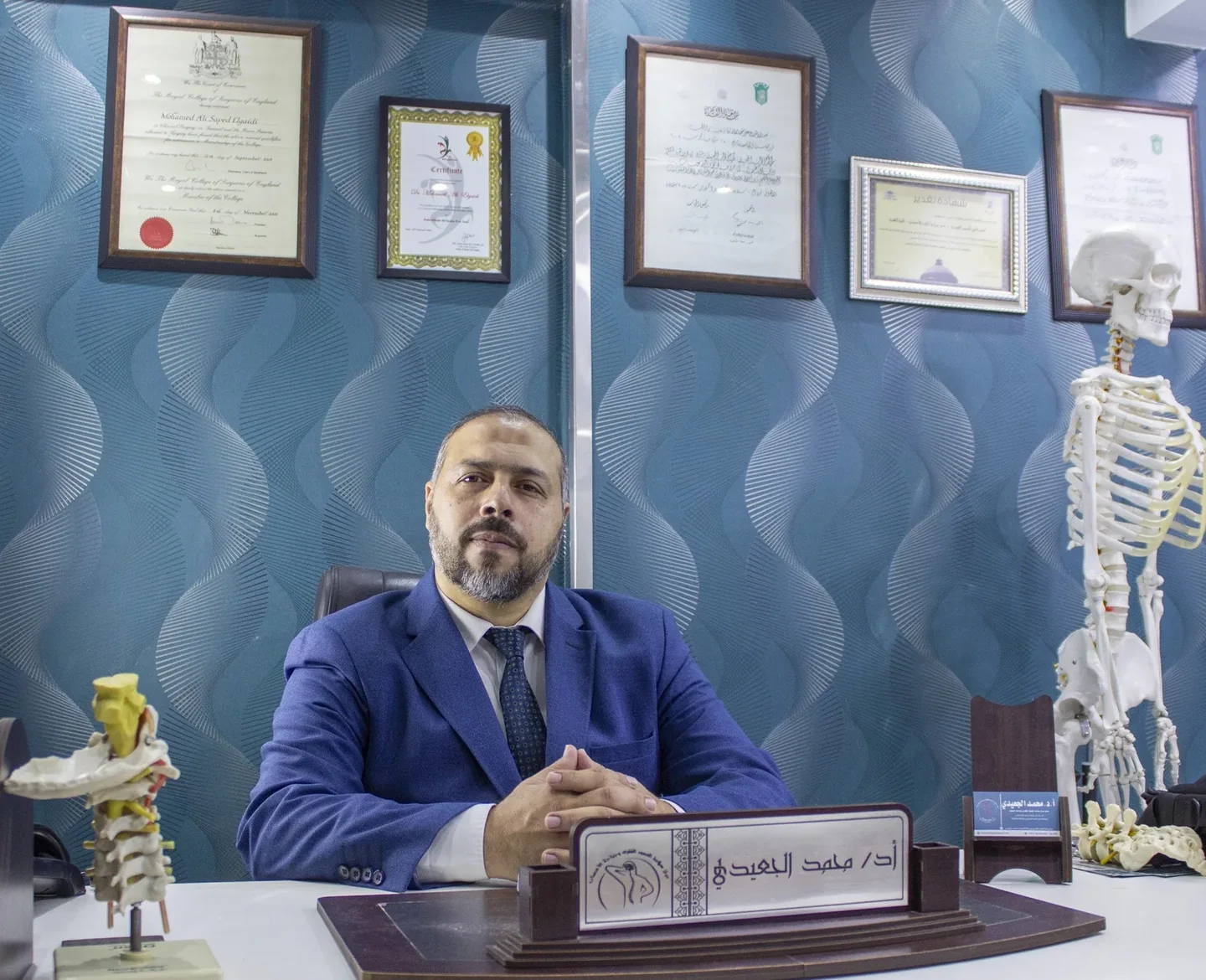
The clinic is at 114 El-Tahrir St., Dokki, Giza, next to Dokki Metro station (4th floor), close to Mohandessin and central Cairo. If you are travelling from elsewhere in Egypt or the Gulf, contact the clinic on WhatsApp before you travel to arrange your appointment. Bring your MRI, X-rays and reports of previous injections and how you responded to them.