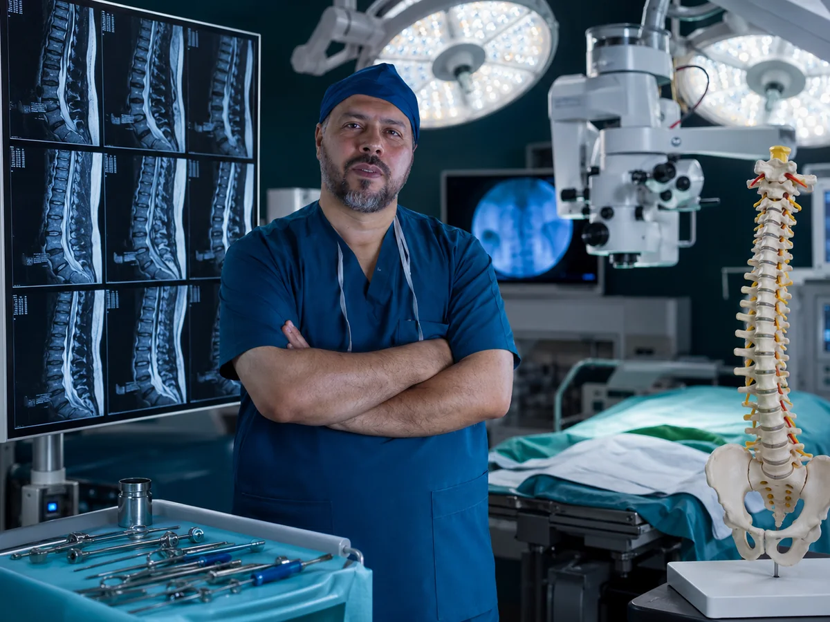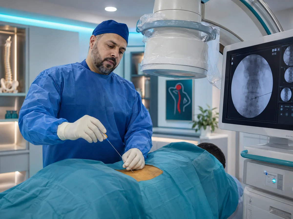
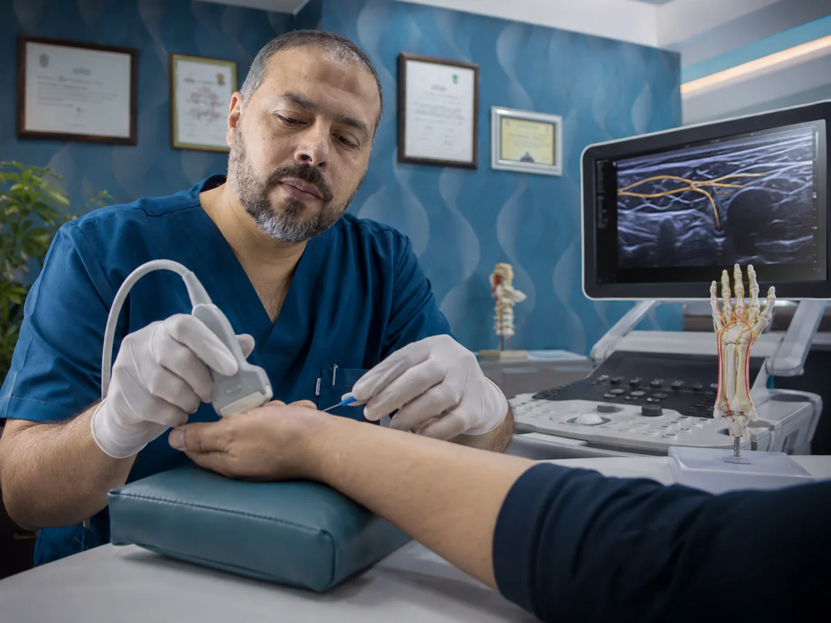
Carpal tunnel surgery releases the median nerve where it is squeezed at the wrist, and it is the most common form of peripheral nerve surgery, the treatment of nerves running from the spinal cord to the arms and legs. At his clinic in Dokki, Cairo, Prof. Mohamed El-Gaidi treats compression, injury and tumours of these nerves, from diagnosis with nerve studies through surgery and rehabilitation.
A peripheral nerve works like a cable carrying both movement and feeling. When it is compressed or cut, symptoms appear in a specific area of the hand or foot, which lets the doctor locate the problem accurately even before any test.
Carpal tunnel syndrome: the numbness that wakes you at night
The carpal tunnel is a narrow passage in the wrist carrying the median nerve and the finger flexor tendons, roofed by a tough ligament. When the space tightens, the nerve is compressed and typical symptoms follow:
- Numbness and tingling in the thumb, index, middle and half the ring finger, with the little finger usually spared.
- Symptoms worse at night that wake you up, eased by shaking the hand.
- Numbness while holding a phone, driving or reading.
- Dropping objects and difficulty with buttons.
- In advanced cases: thumb weakness and wasting of the muscles at its base.
Risk rises with repetitive wrist work, diabetes, an underactive thyroid, rheumatoid arthritis, pregnancy and excess weight. Carpal tunnel can resemble a pinched nerve root in the neck from a cervical disc herniation, and the two may coexist, so the neck is examined too.
Nerve conduction studies: confirming the diagnosis
Diagnosis starts with the examination: sensation in each finger, thumb strength, and simple provocation tests such as tapping or flexing the wrist. Then a nerve conduction study and EMG measures how fast signals travel along the nerve and how the muscles respond.
The study tells us whether compression is present, where it is, and how severe it is, and those answers decide the treatment. Ultrasound may show a swollen nerve inside the tunnel, and MRI is used for tumours and injuries.
When is carpal tunnel surgery needed, and how is it done?
Mild cases often improve with a night splint that keeps the wrist straight, changing strenuous movements, and sometimes a local injection. Surgery is recommended in the following situations:
Numbness persisting despite several weeks of conservative care, thumb weakness or muscle wasting, constant numbness that no longer comes and goes, or moderate to severe compression on the nerve study.
Anaesthesia
The operation is usually done under local anaesthetic to the hand; you stay awake and comfortable.
Releasing the nerve
Through a small incision in the palm, the ligament roofing the tunnel is divided, widening the space and relieving pressure on the median nerve.
Home the same day
The procedure is short, and you go home the same day with a light dressing.
Recovery
You move your fingers from day one, and stitches come out after about two weeks. Grip strength returns gradually over weeks.
Night-time numbness improves quickly for most patients. Long-standing wasting and weakness recover slowly and may not recover fully, which is a clear reason not to postpone surgery once it is needed.
Ulnar nerve entrapment at the elbow
The ulnar nerve is the "funny bone" nerve. It is usually compressed behind the elbow in the cubital tunnel, and less often at the wrist. Symptoms include:
- Numbness in the little finger and half the ring finger, worse with the elbow bent for long periods, such as on the phone or sleeping with the arm flexed.
- Weakness in fine hand movements such as writing or opening jars.
- In advanced cases, wasting between the fingers and clawing of the last two fingers.
Mild cases benefit from not leaning on the elbow and avoiding bending it during sleep. When symptoms persist or weakness appears, ulnar nerve release surgery relieves the pressure. The small hand muscles are sensitive to how long compression lasts, so hand weakness deserves early assessment.
Other trapped nerves in the arm and leg
A nerve can be trapped at any tight point along its course. Examples assessed and treated with nerve entrapment release include:
Radial nerve in the forearm
Forearm pain and weakness lifting the wrist or fingers, sometimes confused with tennis elbow.
Peroneal nerve at the knee
Difficulty lifting the foot (foot drop) and numbness on top of the foot, seen with prolonged leg crossing or after rapid weight loss.
Tarsal tunnel
Burning and numbness in the sole from compression of the nerve behind the inner ankle bone.
Lateral femoral cutaneous nerve
Burning and numbness on the outer thigh, worse with tight belts and weight gain.
Foot drop in particular has several causes, including a compressed nerve root in the lower back, so examination and nerve studies separate a peripheral nerve problem from one that may need spine surgery.
Nerve injuries after cuts and accidents: repair and grafting
Glass and knife wounds, fractures and road accidents can cut, bruise or stretch a nerve. Timing is decisive, because nerves regrow slowly (roughly a millimetre a day) and a muscle that waits too long for its nerve loses its ability to respond over the months.
| Type of injury | Usual timing | Procedure |
|---|---|---|
| Clean, sharp cut through the nerve | Early, within days of injury | Nerve repair with fine sutures joining the two ends |
| Bruising, stretch or injury with a fracture | Follow-up with exams and nerve studies; surgery if no signs of recovery by about three months | Nerve exploration, release or repair |
| A gap between the nerve ends | At repair, or once the wound is ready | Nerve grafting using a segment of another sensory nerve |
| Contaminated or crushed wound | After the wound is cleaned and healed, often within weeks | Delayed repair or grafting |
Nerve grafting is used when the two ends cannot be brought together without tension. The graft is usually taken from a small sensory nerve in the leg, leaving a limited patch of numbness, and acts as a bridge for the nerve to regrow through.
Do not wait: if a cut to your hand, arm or leg is followed by loss of feeling or inability to move the fingers or foot, seek assessment the same day. Early repair improves the chance of recovery.
Brachial plexus injuries
The brachial plexus is the network of nerves leaving the neck to supply the shoulder, arm and hand. It is most often injured in motorcycle accidents and falls onto the shoulder, causing partial or complete paralysis of the arm with loss of sensation.
Assessment relies on careful testing of each muscle, MRI, and nerve studies a few weeks after injury to see which nerves are recovering on their own. Injuries showing no recovery are considered for brachial plexus surgery to explore, repair or graft the nerves, ideally decided within the first months. The aim is usually to restore key functions such as elbow bending and shoulder stability, and recovery takes many months.
Peripheral nerve tumours
A lump under the skin along a nerve that sends tingling into the fingers when tapped may be a nerve tumour. Most are benign. The commonest, the schwannoma, grows from the nerve sheath and can usually be removed while preserving the nerve fibres and function. Neurofibromas are more intertwined with the nerve, and treatment depends on symptoms and size.
Diagnosis is by MRI, sometimes with a biopsy. Any lump that grows quickly, becomes increasingly painful or causes weakness deserves prompt assessment. Peripheral nerve tumour removal calls for a neurosurgeon's experience because the goal is to remove the tumour while protecting nerve function as far as possible.
Rehabilitation after peripheral nerve surgery
Surgery removes the pressure or reconnects the nerve; rehabilitation gets the hand or foot working again. Depending on the case, the plan after surgery includes:
- A splint to protect a repair or prevent joint stiffness in the first weeks.
- Physiotherapy and occupational therapy to restore joint movement and muscle strength.
- Sensory re-education exercises as the nerve recovers.
- Protecting numb skin from burns and cuts until feeling returns.
- Regular follow-up exams, with nerve studies when needed to measure progress.
For persistent nerve pain after an injury, nerve pain medication or guided injections may be added as part of pain management without surgery.
A neurosurgeon following the nerve from diagnosis to rehabilitation
Diagnosing peripheral nerve problems means telling a peripheral nerve from a nerve root in the neck or back, which is the core of neurosurgery and spine surgery. At the Dokki clinic the limb and the spine are examined together, the nerve study is read alongside your symptoms, and the plan and its timing are explained clearly.

Bring any nerve study you have, scans of your neck or the affected limb, previous operation reports and the exact date of your injury, since timing is central to the decision.