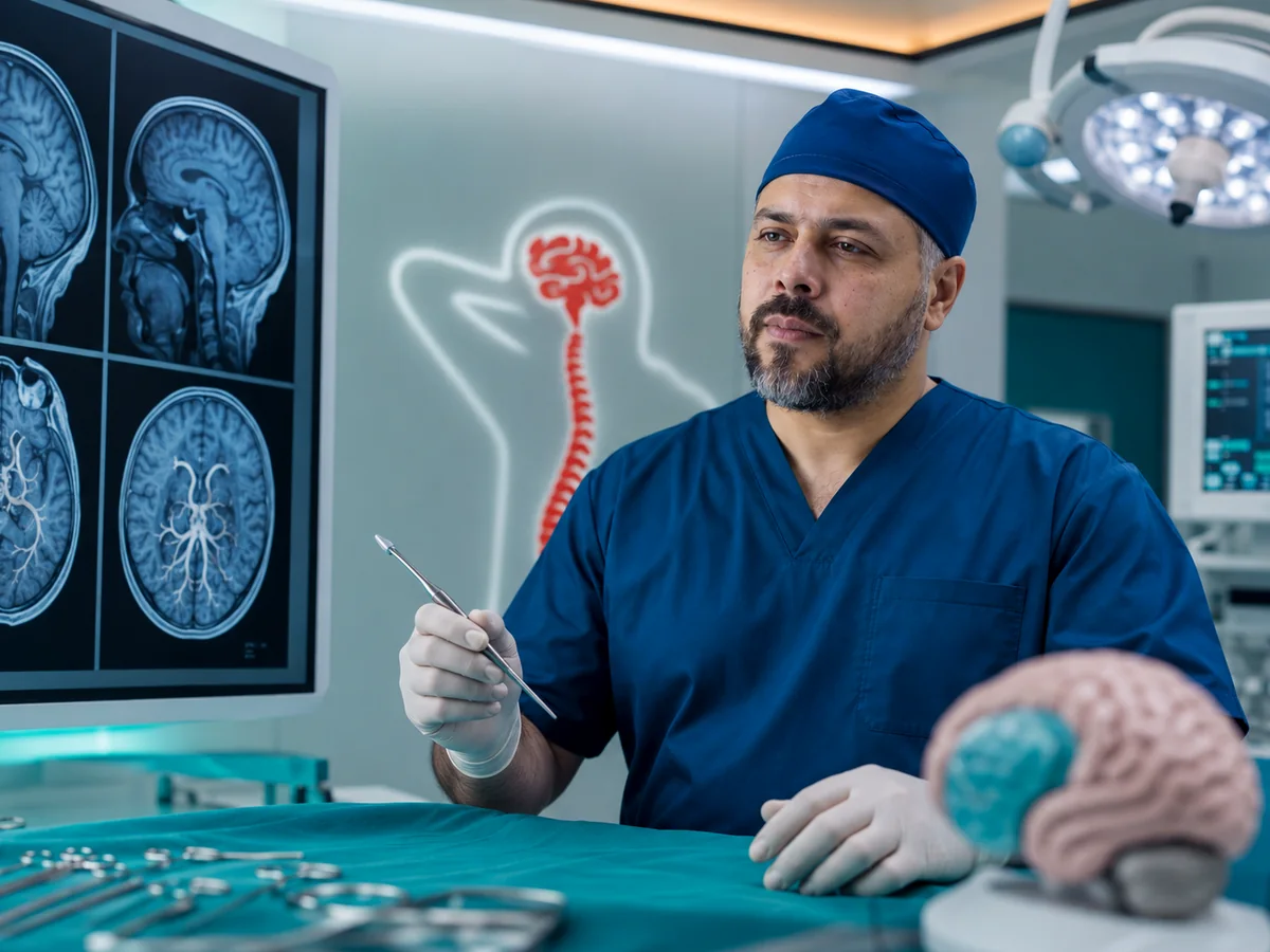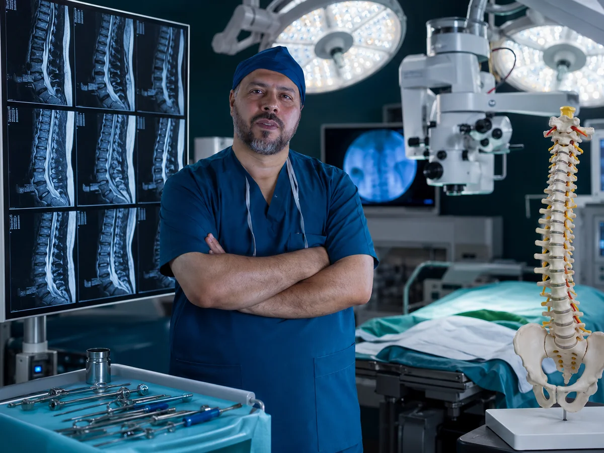
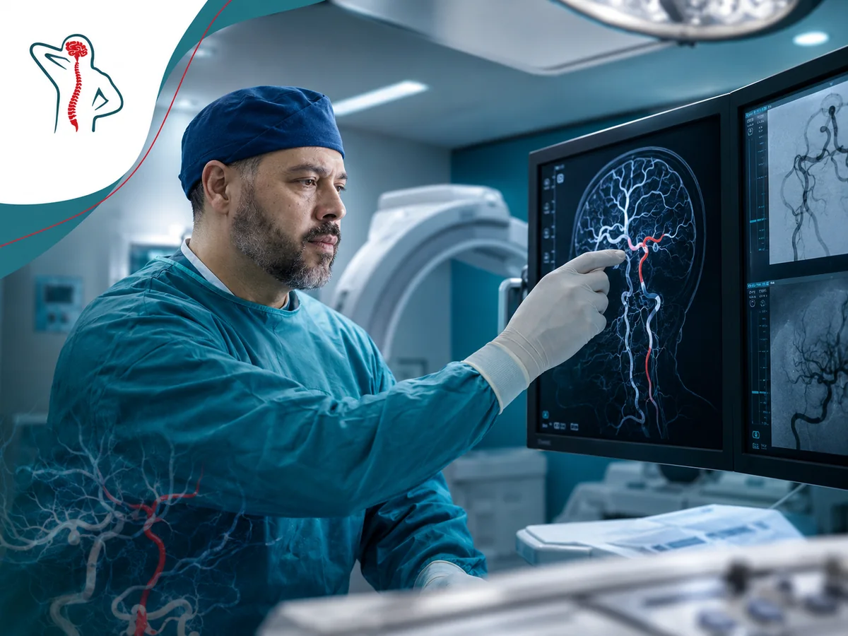
The first step in brain aneurysm treatment is weighing the chance that the aneurysm will bleed against the risks of treating it; every decision follows from that balance. Prof. Mohamed El-Gaidi, Professor of Neurosurgery, sees patients with brain aneurysms, arteriovenous malformations, cavernomas and spinal vascular malformations at his clinic in Dokki, Cairo, and goes through the vascular imaging with them step by step.
A sudden thunderclap headache is an emergency. If a headache hits like an explosion and peaks within seconds, with vomiting, a stiff neck, fainting, a seizure or a drooping eyelid, do not wait for an appointment. Go immediately to the nearest emergency department with a CT scanner.
What is a brain aneurysm?
An aneurysm is a small balloon-like bulge in the wall of a brain artery, usually at a branching point at the base of the brain. Most are small, cause no symptoms and are found by chance on a scan done for headache or dizziness. Smoking, high blood pressure, family history and some inherited conditions raise the risk. The real danger is rupture, which causes bleeding around the brain.
Unruptured aneurysms
Here there is time to think. Future bleeding risk is estimated from size, location, shape, the patient's age and any previous bleed from another aneurysm, then compared with the risk of treatment. Small regular aneurysms are often followed, with blood pressure control and stopping smoking.
Ruptured aneurysms and subarachnoid hemorrhage
When an aneurysm ruptures, blood spreads around the brain: a subarachnoid hemorrhage. CT usually shows the bleed in the first hours, then CT angiography or catheter angiography identifies the aneurysm. Blood pressure is controlled, medication is given to prevent vasospasm, and a temporary drain is placed if acute hydrocephalus develops. The aneurysm is then secured as early as appropriate to prevent a second, often worse bleed, and the patient is monitored in intensive care for days.
AVMs and cavernomas
Arteriovenous malformation (AVM)
A tangle of abnormal vessels where blood passes from arteries to veins without capillaries. Usually present from birth, it may show up as bleeding, seizures, headache or gradual weakness.
Cavernous malformation (cavernoma)
A small mulberry-like cluster of thin-walled, slow-flow vessels. It does not show on angiography but is well seen on MRI. It can cause seizures or small repeated bleeds, and some are familial.
An AVM is assessed by its size, whether it lies in an eloquent brain area and how its veins drain; these decide whether surgery is safe or whether radiosurgery or observation is better. Seizure control with medication, coordinated with a neurologist, is part of treatment whatever is decided. A large blood clot from a bleeding malformation may need urgent evacuation, with the malformation itself treated later.
Spinal vascular malformations
The most common is the spinal dural arteriovenous fistula, typically in men from middle age onward. It causes gradual leg weakness, numbness and bladder problems, and is often first mistaken for spinal stenosis. MRI may show cord swelling with dilated vessels on its surface, and spinal angiography confirms it. Closing the fistula surgically or endovascularly can halt deterioration and improve symptoms, and early treatment makes a difference. For other spinal problems, see spine surgery in Cairo.
Imaging: CTA, MRA and catheter angiography
| Test | What it shows | When it is used |
|---|---|---|
| Non-contrast CT | Bleeding around or inside the brain | First test in the emergency department |
| CT angiography (CTA) | Aneurysm location, size and relation to arteries and bone | Fast in emergencies; useful for surgical planning |
| MR angiography (MRA) | Aneurysms and malformations without X-rays; MRI shows cavernomas | Follow-up of small aneurysms; screening relatives |
| Catheter angiography (DSA) | The most detailed view of vessels and flow, including 3D | Before treatment decisions; AVMs and spinal fistulas |

Clipping, endovascular, radiosurgery or observation?
No single treatment fits every aneurysm or malformation. The decision is discussed between the neurosurgeon and the interventional neuroradiologist, with radiation oncology when relevant, and presented to you with the reasons.
Microsurgical clipping
A small metal clip is placed across the aneurysm neck under the microscope, excluding it from the circulation. Microsurgery is also used to remove AVMs and cavernomas and to evacuate compressing clots.
Endovascular treatment
Fine coils are packed into the aneurysm through a catheter from the groin or wrist, sometimes with a stent. Embolization is also used for AVMs before or instead of surgery.
Radiosurgery
Suits some small, deep AVMs. The AVM closes gradually over years, and the bleeding risk remains until closure is complete.
Observation
A real option for many small aneurysms and asymptomatic cavernomas, with risk-factor control and periodic imaging.
| Comparison | Clipping | Coiling |
|---|---|---|
| Access | Small skull opening | Through the groin or wrist artery |
| Best suited to | Wide-necked aneurysms, branches arising from the sac, or with a compressing clot | Deep and posterior circulation aneurysms; older or frailer patients |
| Stay and recovery | Relatively longer | Usually shorter |
| Durability | Recurrence is rare after complete clipping | Part of the aneurysm may recur and need retreatment |
| Follow-up | Imaging to confirm closure | More regular imaging |
Regular imaging follow-up is a deliberate treatment decision, and sometimes the safest option for you.
Recovery and follow-up
After treatment of an unruptured aneurysm, most patients go home within a few days and resume normal activities within weeks. After subarachnoid hemorrhage recovery is longer: intensive care, vasospasm prevention, sometimes drainage for hydrocephalus, then rehabilitation as needed.
- Control blood pressure and stop smoking completely: the most important things you can do yourself.
- Avoid stimulants and excessive caffeine or energy drinks.
- Moderate exercise such as walking is good; discuss heavy lifting with your doctor.
- Flying is not forbidden with an unruptured aneurysm; discuss pregnancy plans with your neurosurgeon.
- Vascular imaging after treatment, then at intervals set by the type of treatment.
Any change in the pattern of headache, double vision or a drooping eyelid needs urgent contact rather than waiting for the next scan.
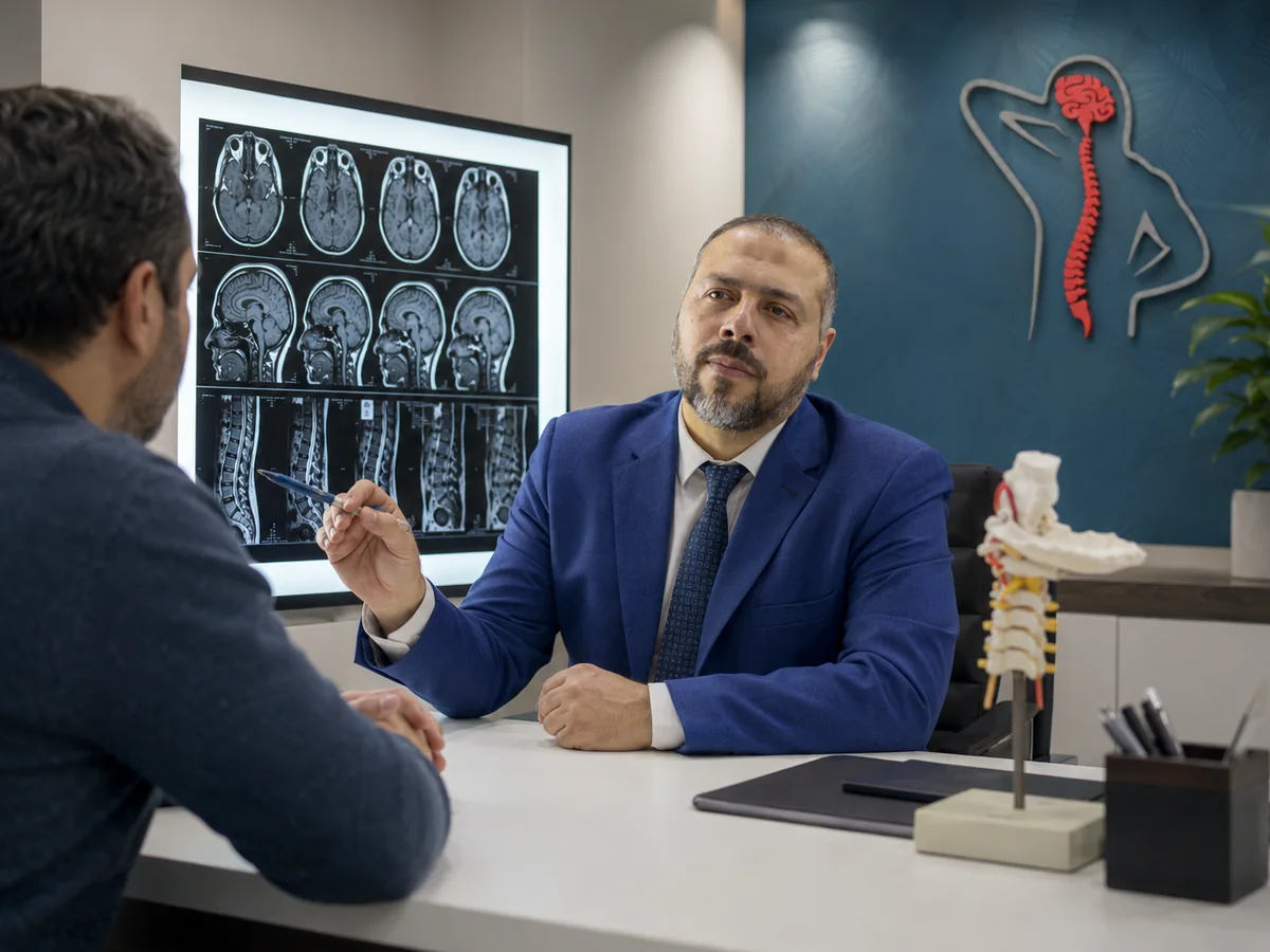
Who will look after you

Cerebrovascular decisions need a surgeon who knows the limits of each treatment, works with interventional radiology and intensive care, and will tell you frankly when observation is enough. Prof. El-Gaidi sees patients at the Spine Health & Neurosurgery Center, 114 El-Tahrir St., Dokki, Giza, next to Dokki Metro station. Bring your CT, MRI and angiography discs and any reports. If a brain bleed was not caused by an aneurysm or malformation, read about brain hemorrhage and brain tumor surgery, or browse all neurosurgery services.