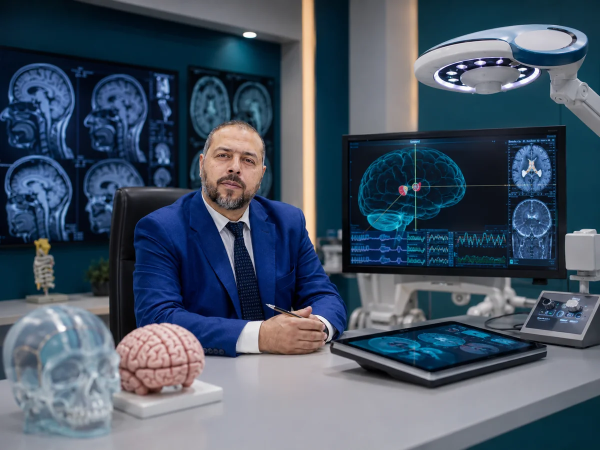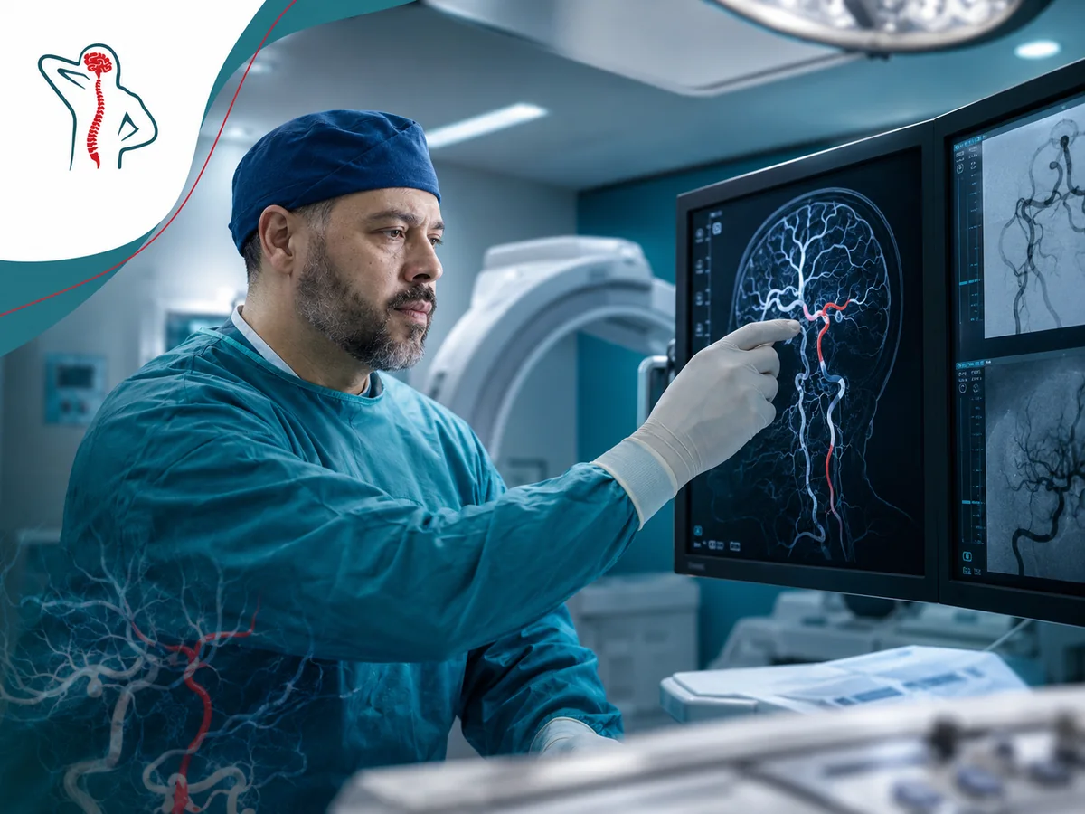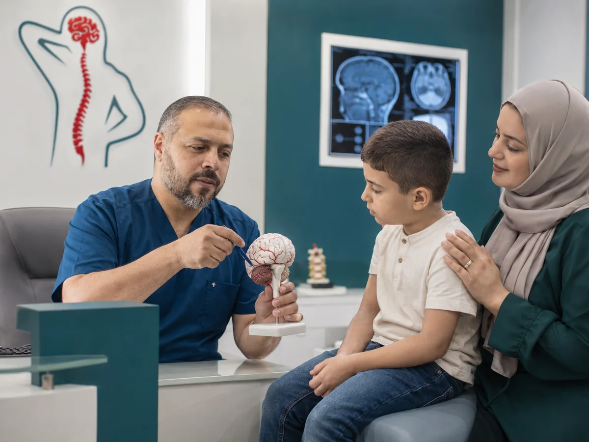
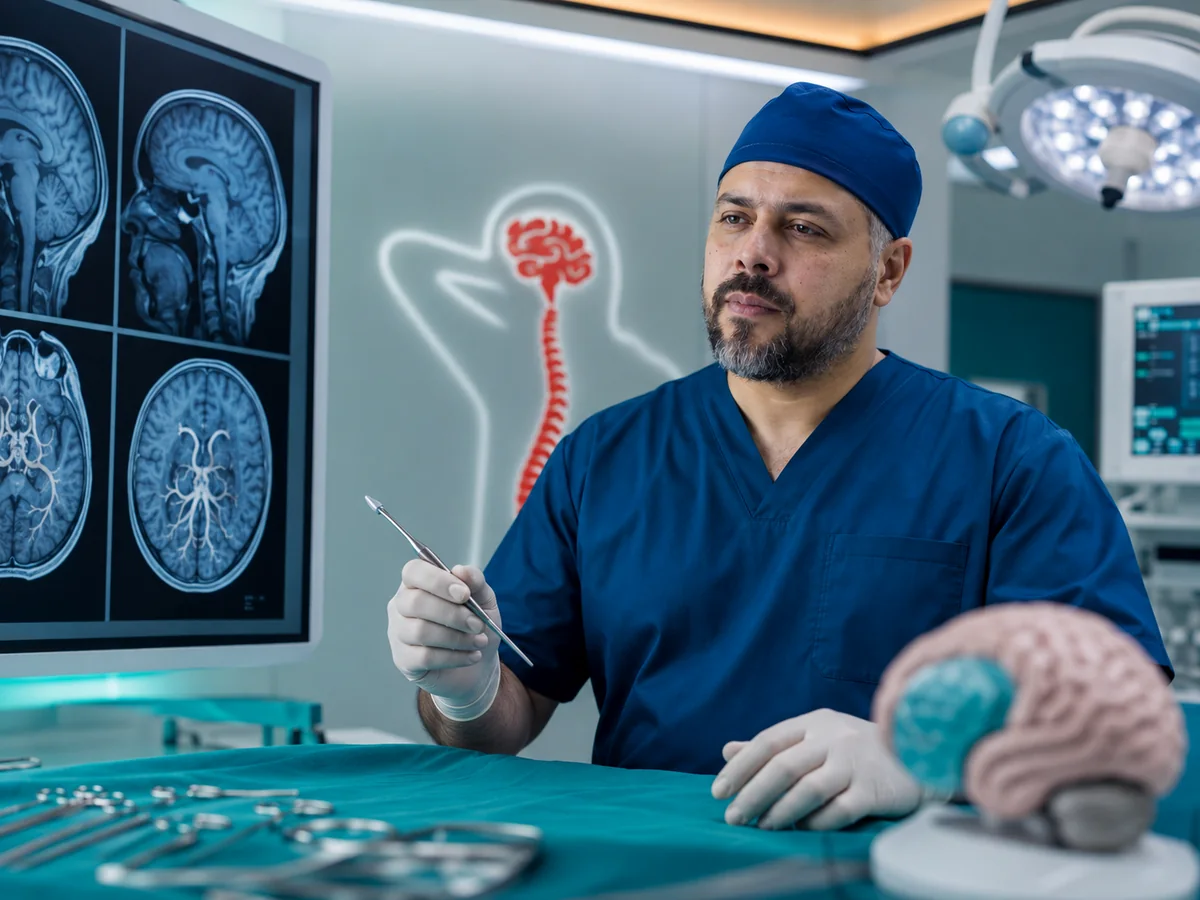
Brain tumor surgery is decided by three questions: what type of tumor this is likely to be, where it sits relative to the areas that control movement, speech and vision, and what removing it will add to the patient's life. Prof. Mohamed El-Gaidi, Professor of Neurosurgery, sees patients with brain, skull base and pituitary tumors at his clinic in Dokki, Cairo, along with brain hemorrhage, blood collections and head injuries.
Warning symptoms of a brain tumor
Symptoms come from pressure on brain tissue or raised pressure inside the skull, so they depend on the tumor's location and growth rate. Some tumors are found by chance on a scan done for something else.
- A new headache that gradually worsens, often worse in the morning or with vomiting.
- A first seizure in an adult.
- Slowly increasing weakness or numbness in an arm or leg.
- Difficulty speaking or understanding, or changes in personality and concentration.
- Blurred vision or loss of side vision.
- Hearing loss in one ear with ringing or imbalance.
- Hormonal changes: stopped periods, milky nipple discharge, enlarging hands and feet.
Go to the emergency department now for sudden drowsiness or confusion, rapidly progressing weakness on one side, a sudden severe headache, seizures that do not stop, or repeated vomiting with sleepiness.
Types of brain tumors in plain language
Meningioma
Arises from the membranes around the brain; mostly benign and slow-growing. Small ones may be watched; large or symptomatic ones are removed, and complete removal can be curative.
Glioma
Grows from supporting cells inside the brain, from low to high grade. Surgery aims for the maximum safe removal and an accurate diagnosis, followed by treatment matched to the grade.
Pituitary adenoma
Usually benign; causes symptoms by pressing on the optic nerves or by disturbing hormones. Some respond to medication; the rest are usually removed endoscopically through the nose.
Skull base tumors
Sit under the brain close to cranial nerves and major arteries, such as some meningiomas and acoustic neuromas. The surgical route needs precise planning.
Metastases
Spread to the brain from cancers elsewhere, such as lung or breast, and are among the most common brain tumors in adults. A single or large symptomatic lesion may be removed, coordinated with radiotherapy.
Ventricular tumors and cysts
May block the flow of brain fluid and cause hydrocephalus; some can be managed endoscopically inside the ventricles.
Diagnosis: contrast MRI and beyond
The key test is a brain MRI with contrast, which shows the size and borders of the tumor, its relation to surrounding structures and the swelling around it. CT shows bleeding, bone and calcification, and is the first scan in emergencies and head injuries. Depending on location, you may also need:
- Pituitary hormone tests.
- Visual field and eye examination.
- Hearing tests for acoustic neuroma.
- Vascular imaging near major arteries.
- Functional MRI or tractography.
- Body scans if a metastasis is suspected.
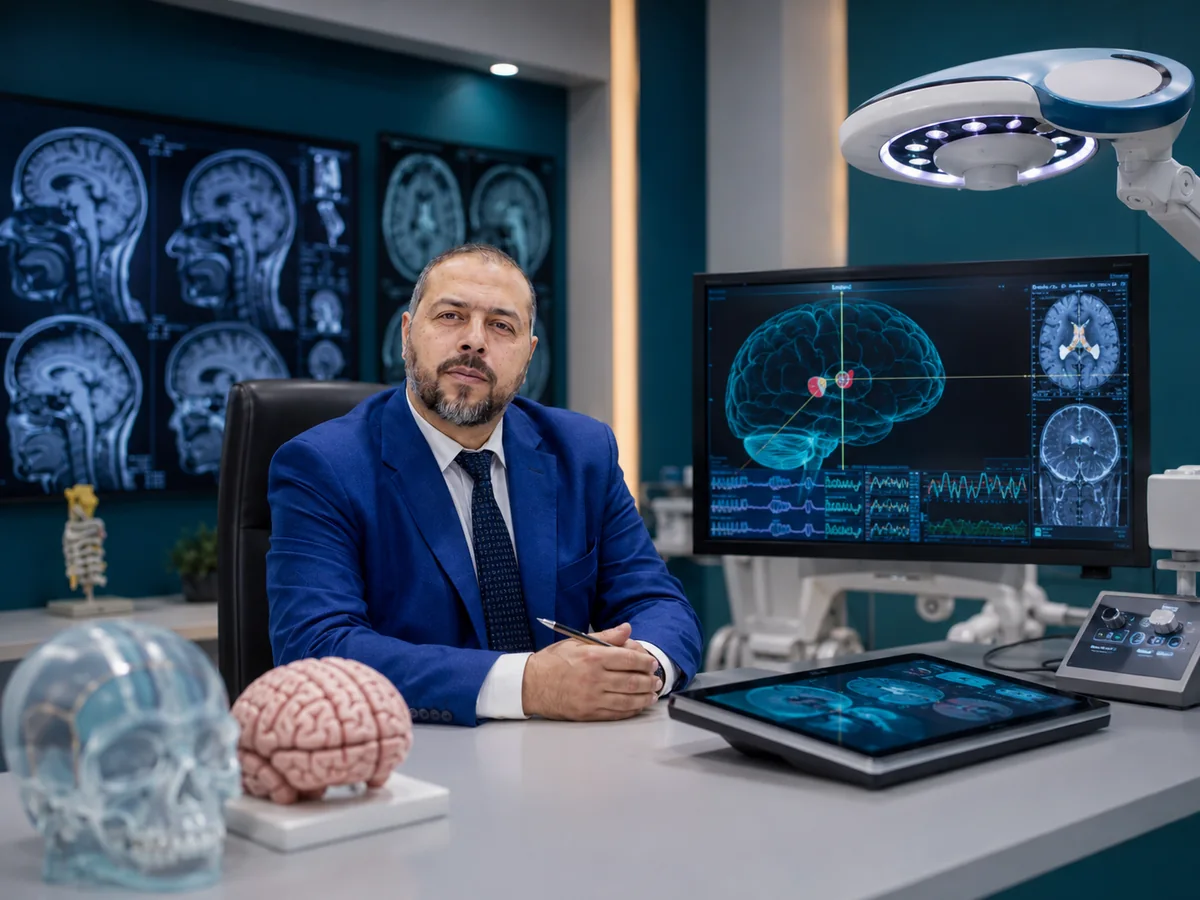
Planning surgery around critical brain areas
Protecting the function of the brain around the tumor comes first. The route to the tumor is mapped on the MRI before surgery, together with its relation to the motor, speech and visual areas and the pathways connecting them. On that basis the aim of surgery is set and explained to you:
- Complete removal when the tumor is well defined and away from critical areas.
- Safe partial removal to relieve pressure when the tumor is attached to areas that cannot be touched, followed by other treatments.
- Biopsy when the tumor is deep or diffuse, to identify it and guide treatment.
At the skull base a small tumor can be harder than a large one on the surface, because of the nerves for vision, hearing, facial movement and swallowing, and the main arteries nearby. The route is chosen to reach the tumor with minimal brain retraction, and a small remnant stuck to a nerve or artery may be left and watched or treated with focused radiation.
Safe removal means taking as much tumor as possible while preserving the patient's ability to move, speak and see.
Preparing for brain surgery
- Blood tests, ECG and an anesthesia assessment, especially with heart disease, diabetes or hypertension.
- Stopping blood thinners for a period your doctor specifies; never stop them on your own.
- Medication to reduce brain swelling, and anti-seizure medication if you have had seizures.
- A recent MRI close to the surgery date for intraoperative planning.
Ask about the exact aim of surgery, the most relevant risks in your case, the expected intensive care stay and when the pathology result will be ready.
Endoscopic transnasal pituitary surgery
The pituitary gland sits in a small bony pocket directly behind the nose, so most pituitary tumors can be reached endoscopically through the nostril, without opening the skull and without a visible scar. Temporary nasal congestion afterwards is common, and you will be asked not to blow your nose forcefully for a while. Urine output and thirst are monitored in the first days, and hormones and vision are rechecked at follow-up, with an endocrinologist when needed.
Brain hemorrhage and blood collections
Not every brain bleed needs an operation. Many hypertensive bleeds are managed in intensive care with blood pressure control. Surgery is considered for large bleeds compressing the brain, cerebellar bleeds, or when the patient deteriorates despite treatment.
Chronic subdural hematoma
Common in older adults and people on blood thinners, blood collects slowly over the brain after a minor bump. Weeks later it shows up as headache, forgetfulness, behavioral change or limb weakness, and is often mistaken for a stroke or dementia. Symptomatic collections are usually drained through small burr holes, most patients improve clearly, and follow-up imaging is needed because it can recur. Bleeds from an aneurysm or vascular malformation are handled differently: see brain aneurysm treatment.
Head injuries and depressed skull fractures
After road accidents or falls, CT shows whether there is an epidural or subdural bleed, a brain contusion or a fracture. An acute compressing bleed is an emergency that needs prompt surgery. A depressed fracture, where a piece of skull is pushed inward, is repaired when it is deep, open or associated with a dural tear, presses on the brain or causes visible deformity. In severe injuries part of the skull may be removed temporarily to relieve pressure on the swollen brain and replaced weeks later.
Recovery and adjuvant therapy
Pathology
The tissue is analyzed to confirm the tumor type and grade, which drives the next step.
Adjuvant treatment
Depending on the result, radiotherapy or chemotherapy is coordinated with oncology.
Rehabilitation
Physiotherapy or speech therapy helps recover affected functions.
MRI follow-up
Regular scans at intervals set by the tumor type, to catch any change early.
Return to desk work is usually after several weeks. Driving is postponed after seizures until your doctor allows it.
Who will look after you

A Kasr Al Ainy graduate with a doctorate in neurosurgery from Cairo University, Prof. El-Gaidi has held the rank of Professor of Neurosurgery since 2023. He sees patients at the Spine Health & Neurosurgery Center, 114 El-Tahrir St., Dokki, Giza, next to Dokki Metro station, easy to reach from Mohandessin and downtown Cairo. Bring your MRI disc or link, older scans and all reports. For children, read about pediatric neurosurgery; if a tumor blocks brain fluid flow, see hydrocephalus treatment. You can also browse all neurosurgery services or read Prof. El-Gaidi's profile.